Why is there a cervical and lumbar enlargement

Sarah Scott
Published Apr 13, 2026
The reason behind the enlargement of the cervical region is because of the increased neural input and output to the upper limbs. An analogous region in the lower limbs occurs at the lumbar enlargement.
Why are the cervical and lumbar enlargements significant?
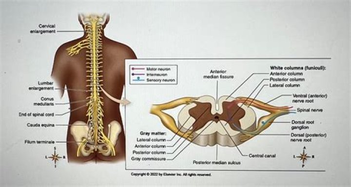
Two regions of the spinal cord are enlarged to accommodate the greater number of nerve cells and connections needed to process information related to the upper and lower limbs (see Figure 1.10B).
Where are the cervical and lumbar enlargements?
Two enlargements of the spinal cord can be visualized: The cervical enlargement, which extends between C3 to T1; and the lumbar enlargements which extends between L1 to S2 (Figure 3.1).
What is the reason for the lumbar enlargement?
The lumbar enlargement of the spinal cord corresponds to the mild increase in cross sectional area of the cord from the T11 level to the conus medullaris. It is enlarged due to the presence of the spinal nerve anterior rami which contribute to the lumbar and sacral plexuses.What is the function of cervical enlargement?
The cervical and lumbar regions of the spinal cord show a larger diameter compared to the rest of the regions. The cervical enlargement and lumbar enlargement represent an increased amount of neurons in the gray matter and axons in the white matter that serve the upper limbs and lower limbs, respectively.
Is Spinal stenosis a degenerative disease?
Lumbar spinal stenosis is a narrowing of the spinal canal, compressing the nerves traveling through the lower back into the legs. While it may affect younger patients, due to developmental causes, it is more often a degenerative condition that affects people who are typically age 60 and older.
What is the pia mater?
The pia mater is the meningeal envelope that firmly adheres to the surface of the brain and spinal cord. It is a very thin membrane composed of fibrous tissue covered on its outer surface by a sheet of flat cells thought to be impermeable to fluid.…
What is the function of the cervical enlargement of the spinal cord quizlet?
The cervical enlargement of the spinal cord is the source of the spinal nerves that contribute to the brachial plexus and supply the upper limbs.How long is the lumbar enlargement?
The lumbar enlargement is 8 cm long from T9 to T12 and in continuity with the conus medullaris, which tapers off at the level of the L1–2 disc space into the filum terminale, an atrophic remnant of the caudal segment of the embryonic spinal cord.
What is the function of cauda equina?The cauda equina is the sack of nerve roots (nerves that leave the spinal cord between spaces in the bones of the spine to connect to other parts of the body) at the lower end of the spinal cord. These nerve roots provide the ability to move and feel sensation in the legs and the bladder.
Article first time published onWhat does the sacral plexus do?
The sacral plexus (plexus sacralis) is a nerve plexus that provides motor and sensory nerves for the posterior thigh, most of the lower leg, the entire foot, and part of the pelvis (see the following image).
What is the lumbosacral plexus?
The lumbosacral plexus is a network of nerves derived from lumbar and sacral roots with each one of them dividing into anterior and posterior branches. … The anterior branches supply the flexor muscles of thigh and leg and posterior branches supply the extensor and abductor muscles.
Which spinal segment of cervical enlargement of spinal cord shows maximum circumference?
The greatest circumference of the enlargement is at the C6 level, which is approximately 38 mm. It extends from about the third to the fifth cervical to the first thoracic vertebra reaching a maximum circumference of about 38 mm.
Where would a doctor perform a lumbar puncture spinal tap on a patient?
A lumbar puncture (spinal tap) is performed in your lower back, in the lumbar region. During a lumbar puncture, a needle is inserted between two lumbar bones (vertebrae) to remove a sample of cerebrospinal fluid.
Where is the lumbar plexus?
It is located on the posterior abdominal wall, anterior to the transverse processes of the lumbar vertebrae and within the posterior portion of the psoas major muscle. The lumbar plexus gives rise to several branches which supply various muscles and regions of the posterior abdominal wall and lower limb.
Is a medulla oblongata?
Medulla oblongataSection of the medulla oblongata at about the middle of the olivary bodyDetailsPart ofBrain stemIdentifiers
What is the GREY matter?
Anatomical terminology. Grey matter (or gray matter) is a major component of the central nervous system, consisting of neuronal cell bodies, neuropil (dendrites and unmyelinated axons), glial cells (astrocytes and oligodendrocytes), synapses, and capillaries.
Can the pia mater be removed?
CSF and the meninges are designed to protect the brain and spinal cord from mechanical shock. … The pia mater is very delicate and adheres to the brain’s surface tenaciously; it will take considerable patience and care to remove it.
What are the final stages of spinal stenosis?
Spinal stenosis, often an end stage of the spine degenerative process, is characterized by leg pain with walking. Pain will go away with rest but you may have to specifically sit down to ease the leg pain.
What is the surgery for cervical spinal stenosis?
Examples of surgical procedures to treat spinal stenosis include: Laminectomy. This procedure removes the back part (lamina) of the affected vertebra. A laminectomy is sometimes called decompression surgery because it eases the pressure on the nerves by creating more space around them.
What are the symptoms of cervical spinal stenosis?
Symptoms usually develop gradually over a long period of time and may include: Stiffness, pain, numbness, or weakness in the neck, shoulders, arms, hands, or legs. Balance and coordination problems, such as shuffling or tripping while walking. Cervical spinal stenosis can be crippling if the spinal cord is damaged.
Which spinal nerves affect which parts of the body?
The nerves of the cervical spine go to the upper chest and arms. The nerves in your thoracic spine go to your chest and abdomen. The nerves of the lumbar spine then reach to your legs, bowel, and bladder. These nerves coordinate and control all the body’s organs and parts, and let you control your muscles.
How many cervical spinal nerves are there?
There are 31 pairs of spinal nerves and roots. Eight pairs of cervical nerves exit the cervical cord at each vertebral level. One member of the pair exits on the right side and the other exits on the left. The first cervical root exits above the C1 vertebra.
What part of the spinal cord carries motor info from the brain?
The anterior root is the motor (efferent) root that carries motor information to the body from the brain. The spinal nerve emerges from the spinal column through the opening (intervertebral foramen) between adjacent vertebrae.
What does the cervical enlargement innervate?
The cervical enlargement is the site of the cell bodies of the motor neurons that innervate the upper limbs, as well as the site where the sensory nerves from the upper limbs synapse.
What do the cervical and brachial plexuses have in common?
What do the cervical and brachial plexuses have in common? Both plexuses contain ventral rami of C5 spinal nerves. The brachial plexus is more complex than the cervical plexus. … The sciatic nerve, formed of ventral rami, is the largest nerve in the human body.
What is the significance of the Conus Medullaris?
The conus medullaris give rise to the lumbar sympathetic, sacral somatic and sacral parasympathetic nerves which continue downward within the cauda equina. These nerves have important functions which can be impaired by injury or ischemia.
What are the first signs of cauda equina?
- Lower limb weakness and intermittent changes in sensation, such as numbness.
- “Saddle anesthesia” – loss or diminished sensation in areas where a person would sit on a saddle.
- Urinary and/or bowel problems, such as retention or incontinence.
What is the most common cause of cauda equina syndrome?
- A severe ruptured disk in the lumbar area (the most common cause)
- Narrowing of the spinal canal (stenosis)
- A spinal lesion or malignant tumor.
- A spinal infection, inflammation, hemorrhage, or fracture.
What is the most common finding in cauda equina syndrome?
Saddle and perineal hypoesthesia or anesthesia. Bowel and bladder disturbances. Lower extremity motor weakness and sensory deficits. Reduced or absent lower extremity reflexes.
What are the cervical spinal nerves?
Cervical spinal nerves, also called cervical nerves, provide functional control and sensation to different parts of the body based on the spinal level where they branch out from the spinal cord.