What muscle compresses abdominal contents

Ava Wright
Published Apr 03, 2026
The transversus abdominis muscle
Which muscle flexes the vertebral column and compresses the abdomen?
Name of MuscleActionExternal abdominal obliqueCompression of abdomen, flexion and rotation of vertebral columnInternal abdominal obliqueCompression of abdomen, flexion and rotation of vertebral columnTransversus abdominisCompression of abdomenRectus abdominisCompression of abdomen and flexion of vertebral column
What are the upper abdominal muscles?
Your abdominal muscles have many important functions, from holding organs in place to supporting your body during movement. There are five main muscles: pyramidalis, rectus abdominus, external obliques, internal obliques, and transversus abdominis.
What are the 4 abdominal muscles?
Muscles of the Abdomen These muscles of the anterolateral abdominal wall can be divided into four groups: the external obliques, the internal obliques, the transversus abdominis, and the rectus abdominis (Figure 16.16 and Table 16.6). Figure 16.16.What is rectus abdominis muscle?
The rectus abdominis muscles are a pair of long, straight muscles that flex the spine and tighten the intra-abdominal wall. They arise from the symphysis pubis and the pubic crest and insert on the linea alba and at the fifth, sixth, and seventh costal cartilages.
What is included in the core muscles?
The major muscles of your core include your transverse abdominis, multifidus, internal and external obliques, erector spinae, diaphragm, pelvic floor muscles, and (of course) your abs, the rectus abdominis. Your minor core muscles include your lats, traps, and (to the surprise of many people) your glutes.
What are the three layers of abdominal muscles?
In medical vernacular, the term ‘abdominal wall’ most commonly refers to the layers composing the anterior abdominal wall which, in addition to the layers mentioned above, includes the three layers of muscle: the transversus abdominis (transverse abdominal muscle), the internal (obliquus internus) and the external …
Which type of muscle is present in abdominal wall?
abdominal muscle, any of the muscles of the anterolateral walls of the abdominal cavity, composed of three flat muscular sheets, from without inward: external oblique, internal oblique, and transverse abdominis, supplemented in front on each side of the midline by rectus abdominis.What are the side abdominal muscles called?
Your oblique muscles (side abdominals) help you bend from the side or twist your torso. Strong obliques support the lower back, warding off back pain and posture problems. Improved posture, thanks to strong obliques, slims your waist.
Which muscle compresses the abdominal viscera during forced expiration?OriginPubic symphysis, pubic crestBlood supplyInferior epigastric and superior epigastric arteries; contributions from posterior intercostal, subcostal and deep circumflex arteriesFunctionTrunk flexion, compresses abdominal viscera, expiration
Article first time published onWhat are the 4 layers of abdominal muscles going from superficial to deep?
- Skin.
- Subcutaneous tissues (further divided into the more superficial Camper’s fascia and the deeper Scarpa’s fascia)
- External oblique muscle.
- Internal oblique muscle.
- Transversus abdominis muscle.
What are the 6 pack muscles?
The term “six-pack’” typically refers to the rectus abdominis muscle. This long, relatively narrow muscle runs from your sternum to your pubic bone and is responsible for dynamically flexing your spine forward ( 1 ).
What is abdominis or abdominus rectus?
The rectus abdominis muscle, also known as the “abdominal muscle” or simply the “abs”, is a paired muscle running vertically on each side of the anterior wall of the human abdomen, as well as that of some other mammals.
What is arcuate line?
The arcuate line is the inferior margin of the posterior leaflet of the rectus sheath within the abdomen. The posterior leaflet of the sheath is formed, superficial to deep, from the: posterior part of the internal oblique aponeurosis. transversus abdominis aponeurosis.
Is the trunk midline muscle that compresses the abdomen?
Linea alba The is the trunk midline muscle that compresses the abdomen. … Quadratus lumborum The is a fibrous line located along the midline of the trunk. Rectus abdominis is a superficial lateral muscle of the abdomen. Internal intercostal External intercostal is a neck muscle that extends the head.
What are the 5 layers of abdomen?
- Mucosa. This is the first and innermost layer or lining. …
- Submucosa. This second layer supports the mucosa. …
- Muscularis. The third layer is made of thick muscles. …
- Subserosa. This layer contains supporting tissues for the serosa.
- Serosa. This is the last and outermost layer.
What is internal oblique?
Internal oblique. Internal abdominal oblique is a muscle found on the lateral side of the abdomen. It is broad and thin. it forms one of the layers of the lateral abdominal wall along with external oblique on the outer side and transverse abdominis on the inner side. Its fibers are obliquely oriented hence the name.
Is latissimus dorsi a core muscle?
Latissimus Dorsi is often thought of as a shoulder muscle, but it has a major role in trunk movement. Having strong core muscles can prevent low back problems and enhances performance. …
How many muscles are in the core?
The Core is composed of as many as 35 different muscle groups connecting into the pelvis from the spine and hip area. In order to simplify the Core muscles I have divided them into four regions; back extensors, abdominals, lateral trunk muscles, and the hip muscles.
Is the core only abdominal muscles?
What you probably don’t realize is that your core is so much more than abdominal muscles. It’s actually made up of 29 muscles in your mid and low back, pelvic floor, butt, and hips. Together, these muscles act as the cornerstone for all of your body’s movements.
What muscle is your oblique?
Oblique muscle refers to two abdominal muscles – the external and internal obliques. These provide trunk flexion and rotation. The external oblique is the thickest and runs from the lower ribs to the iliac crest.
What muscles are in your left side?
- Superficial muscles of left shoulder. Left trapezius. Left deltoid. Left latissimus dorsi.
- Muscles of left rotator cuff. Left infraspinatus. Left teres minor. Left supraspinatus. Left subscapularis.
- Deep muscles of left shoulder. Left levator scapulae. Left rhomboid minor. Left rhomboid major. Left serratus anterior.
What are the trunk muscles?
There are five muscles that form the abdominal part of the anterior trunk. These are the rectus abdominis, pyramidalis, external abdominal oblique, internal abdominal oblique and transversus abdominis. The first three are classified as vertical muscles and they are located near the midline.
Which muscles are found in the walls of stomach and intestine?
Smooth muscles are found in the walls of stomach and intestines.
Are smooth muscles present in abdominal wall?
Smooth muscle is found in the walls of hollow organs, including the stomach, intestines, bladder and uterus; in the walls of passageways, such as blood, and lymph vessels, and in the tracts of the respiratory, urinary, and reproductive systems.
Which type of muscle is present in Iris?
The iris consists of two sheets of smooth muscle with contrary actions: dilation (expansion) and contraction (constriction). These muscles control the size of the pupil and thus determine how much light reaches the sensory tissue of the retina.
Is rectus abdominis superficial to external oblique?
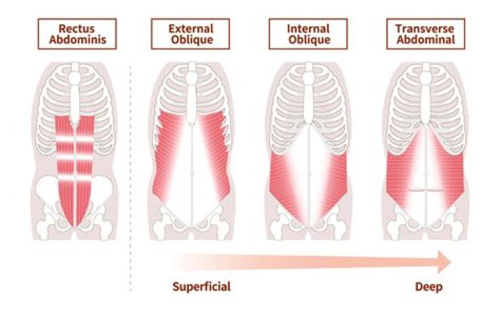
Muscles of the anterior abdominal wall consists of two vertical muscles located on the midline and bisected by linea alba; Rectus abdominis and pyramidalis and three flat muscles on the anterolateral side arranged from superficial to deep; external abdominal oblique, internal abdominal oblique, transversus abdominis.
What is the most superficial muscle of the abdominals Issa?
External Oblique – the most superficial and also the largest flat muscle of the abdominal wall. It runs in an inferior-medial direction and at the midline, its fibers form an aponeurosis and in the midline merge with the linea alba.
What is the fascia Transversalis?
The transversalis fascia is a thin layer of connective tissue lining most of the abdominal cavity between the posterior surface of the transversus abdominis and superficial to the extraperitoneal fat and peritoneum.
What are the contents of rectus sheath?
The rectus sheath is the durable, resilient, fibrous compartment that contains both the rectus abdominis muscle and the pyramidalis muscle. The fascial coverings of the external oblique, internal oblique, and transversus abdominis muscles comprise the rectus sheath.
Which muscle do you contract when you compress the anterior abdominal wall quizlet?
When the internal obliques and transversus abdominis contract, the thoracolumbar fascia is pulled on, compressing the back muscles.