What are the parts of the hippocampus

John Castro
Published Apr 01, 2026
Hippocampus contains two parts: Cornu ammonis (hippocampus proper) and dentate gyrus. Both of these parts are separated by hippocampal sulcus and curve into each other. Below the sulcus lies subiculum. Since hippocampus is a part of allocortex (archicortex), there is a zone that separates it from neocortex.
What 3 areas are controlled by the hippocampus?
Being an integral part of the limbic system, hippocampus plays a vital role in regulating learning, memory encoding, memory consolidation, and spatial navigation.
How many hippocampus are in the brain?
Because the brain is lateralized and symmetrical, you actually have two hippocampi. They are located just above each ear and about an inch-and-a-half inside your head.
What are the different regions of the hippocampus?
Anatomical terms of neuroanatomy The hippocampus proper refers to the actual structure of the hippocampus which is made up of three regions or subfields. The subfields CA1, CA2, and CA3 use the initials of cornu Ammonis, an earlier name of the hippocampus.Is the amygdala part of the hippocampus?
amygdala, region of the brain primarily associated with emotional processes. … The amygdala is located in the medial temporal lobe, just anterior to (in front of) the hippocampus. Similar to the hippocampus, the amygdala is a paired structure, with one located in each hemisphere of the brain.
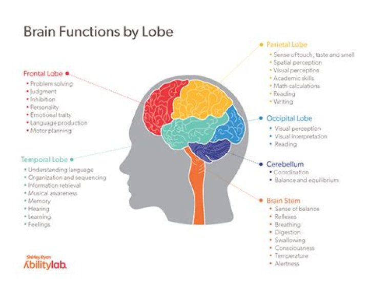
What lobe is the hippocampus in?
Hippocampus is a complex brain structure embedded deep into temporal lobe. It has a major role in learning and memory.
What are the main functions of the hippocampus?
The hippocampus is thought to be principally involved in storing long-term memories and in making those memories resistant to forgetting, though this is a matter of debate. It is also thought to play an important role in spatial processing and navigation.
What types of neurons are in the hippocampus?
Neurons in cortical areas, including the hippocampus, can be broadly divided into two major classes: principal cells and non-principal cells or interneurons. Prin- cipal cells comprise the majority (∼80–90%) of the neuronal population and show largely homogeneous but area-specific morphological features.What surrounds the hippocampus?
The parahippocampal gyrus (or hippocampal gyrus) is a grey matter cortical region of the brain that surrounds the hippocampus and is part of the limbic system. The region plays an important role in memory encoding and retrieval.
What type of cells are in the hippocampus?The stratum oriens layer contains two types of cells: basal dendrites and basket cells. The pyramidal cell layer, consistent with its name, contains pyramidal cells of the hippocampus.
Article first time published onWhy hippocampus is called Seahorse?
Seahorses are scientifically classified in the family Syngnathidae, a name that comes from the Greek words for “jaw” and “together.” Hippocampus, which comes from the Greek words for “horse” and “sea animal,” is the genus that seahorses are classified as.
How many neurons are in the human hippocampus?
Within the cerebral cortex, the elephant hippocampus weighs 24.42 g and has a slightly larger volume than the human hippocampus (Patzke et al., 2013), but holds only 36.63 million neurons bilaterally, compared to approximately 250 million neurons in the ensemble of the human hippocampus plus amygdala (Andrade-Moraes et …
Where are memories stored?
The hippocampus, located in the brain’s temporal lobe, is where episodic memories are formed and indexed for later access. Episodic memories are autobiographical memories from specific events in our lives, like the coffee we had with a friend last week.
What part of brain controls fear and anxiety?
The amygdala is responsible for the expression of fear and aggression as well as species-specific defensive behavior, and it plays a role in the formation and retrieval of emotional and fear-related memories.
What parts of the brain are used for memory?
Most available evidence suggests that the functions of memory are carried out by the hippocampus and other related structures in the temporal lobe. (The hippocampus and the amygdala, nearby, also form part of the limbic system, a pathway in the brain (more…)
Is the hippocampus part of the limbic system?
The limbic system includes the hippocampal formation, amygdala, septal nuclei, cingulate cortex, entorhinal cortex, perirhinal cortex, and parahippocampal cortex. These last three cortical areas comprise different portions of the temporal lobe. … Hippocampus means seahorse in Greek.
What emotions does the hippocampus control?
The hippocampus, located in the medial temporal lobe and connected with the amygdala that controls emotional memory recalling and regulation (Schumacher et al., 2018); it has increased the functional connectivity with anterior cingulate or amygdala during emotional regulation and recalling of positive memory (Guzmán- …
What techniques are used to view the hippocampus?
Since the early 1990’s, Magnetic Resonance Imaging (MRI) has been used to produce accurate hippocampal volume measurements, by separating hippocampal structures not only from the surrounding white matter (WM), but also from contiguous areas of gray matter (GM).
Can you live without a hippocampus?
In short, the hippocampus orchestrates both the recording and the storage of memories, and without it, this “memory consolidation” cannot occur.
What can damage hippocampus?
Damage to hippocampus can occur through many causes including head trauma, ischemia, stroke, status epilepticus and Alzheimer’s disease.
What is the major output of the hippocampus?
The major input to the hippocampus is through the entorhinal cortex (EC), whereas its major output is via CA1 to the subiculum. Information reaches CA1 via two main pathways, direct and indirect.
What connects hippocampus and hypothalamus?
Fornix: an arching, band of white matter axons (nerve fibers) that connect the hippocampus to the hypothalamus.
What artery supplies the hippocampus?
Arterial vascularization of the hippocampus is dependent on the collateral branches of the posterior cerebral artery and the anterior choroidal artery, forming the network of superficial hippocampal arteries that in turn lead to deep intrahippocampal arteries.
What are hippocampal neurons?
Hippocampal neurons play a major role in the functioning of the human brain. … The hippocampus belongs to the limbic system and plays an important role in the consolidation of information from short to long-term memory, and enables navigation via spatial memory.
What neurotransmitter does the hippocampus release?
Major neurotransmitter distinctions Hippocampal neurons mainly release glutamate or gamma-aminobutyric acid (GABA) (Kullmann, 2007).
What are sensory neurons?
Sensory neurons are the nerve cells that are activated by sensory input from the environment – for example, when you touch a hot surface with your fingertips, the sensory neurons will be the ones firing and sending off signals to the rest of the nervous system about the information they have received.
Is the hippocampus an organ?
Your brain forms, organizes, and stores memories in the hippocampus. This tiny organ helps you form long-term memories, connect memories to other memories, and connect memories to emotions and senses. … The organ consists of two parts: one on the right side of your brain and one on the left.
Do male seahorses get pregnant?
Seahorses and their close relatives, sea dragons, are the only species in which the male gets pregnant and gives birth. Male seahorses and sea dragons get pregnant and bear young—a unique adaptation in the animal kingdom. Seahorses are members of the pipefish family.
Can you eat seahorse?
Unfortunately seahorses are edible, and as regarded as a delicacy in China, Japan and other Asian countries. The demand has put great risk of extinction on these timid creatures.
What do Hippocampus kuda eat?
- fish.
- aquatic or marine worms.
- aquatic crustaceans.
- zooplankton.
How many brains do elephants have?
Total cortical surface area, pial257,067 mm2Exposed cortical surface area62,146 mm2Average folding index4.18