Is the obturator nerve a peripheral nerve

Ethan Hayes
Published Apr 12, 2026
The obturator nerve is a major peripheral nerve of the lower limb.
What group of nerves does the obturator nerve belong to?
The obturator nerve is one of the largest branches of the lumbar plexus. It is a mixed nerve which arises from the ventral (anterior) rami of the spinal nerves L2-L4. Motor: Adductor longus, adductor brevis, gracilis, obturator externus and ischiocondylar part of adductor magnus muscle.
What forms the obturator nerve?
The obturator nerve arises from the anterior division of the L2–L4 nerve roots and courses through the psoas major before emerging from the medial border to predominantly innervate the thigh adductor muscles.
What is a obturator nerve?
The obturator nerve arises from the lumbar plexus and provides sensory and motor innervation to the thigh. This nerve provides motor innervation to the medial compartment of the thigh and as a result, is essential to the adduction of the thigh.What is the difference between femoral nerve and obturator nerve?
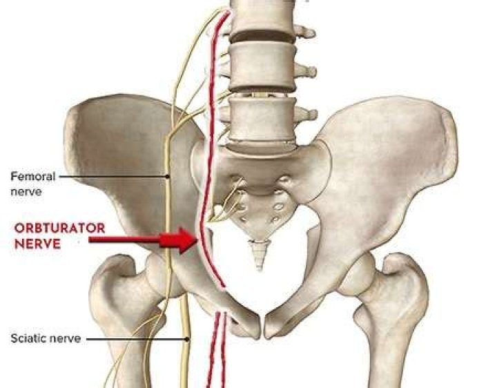
Both these nerves arise from the lumbar plexus, which lies up here within the thickness of the psoas major muscle. The femoral nerve emerges lateral to psoas major, the obturator nerve medial to it. … It runs across the iliacus muscle, and passes under the inguinal ligament just lateral to the femoral artery.
Is obturator nerve motor or sensory?
The obturator nerve is derived from L2-4 and travels along the medial border of the psoas muscle; it is both a motor and a sensory nerve. It travels through the obturator foramen with the obturator artery and vein into the thigh. The obturator nerve divides into anterior and posterior branches.
Between what two muscles is the obturator nerve?
Descends in a plane between the adductor longus and adductor brevis (towards the femoral artery). Here, it supplies motor fibres to the adductor longus, adductor brevis and gracilis. It can also supply the pectineus muscle. It then pierces the fascia lata to become the cutaneous branch of the obturator nerve.
What is obturator canal?
The obturator canal is a small opening in the superior aspect of the obturator foramen that connects the pelvis to the medial compartment of the thigh. The obturator foramen is otherwise covered by the obturator membrane.What is the purpose of the obturator?
The obturator is used to insert a tracheostomy tube. It fits inside the tube to provide a smooth surface that guides the tracheostomy tube when it is being inserted.
Where does the obturator nerve split?The nerve path continues by following along the lateral wall of the pelvis, passing through the obturator canal, to enter the medial compartment of the thigh. From here the nerve divides into the anterior and posterior branch which are separated by the adductor brevis muscle.
Article first time published onHow do you test the obturator nerve?
Perhaps the best test to confirm obturator neuropathy is needle electromyography (EMG). Bradshaw et al. showed chronic denervation in the short and long adductor muscles of athletes with chronic groin pain attributed to obturator neuropathy [4].
Where is obturator artery?
Branching from the internal iliac artery, the obturator artery runs a course along the pelvic wall. It runs to the upper portion of the obturator foramen, which is an opening for blood vessels and nerves between the ischium and pubis bones, located in the lower part of the pelvis.
What are the signs and symptoms of obturator nerve entrapment?
- reduced range of movement.
- swelling/inflamation.
- stiffness.
- weakness.
- numbness.
- spasm.
What nerve runs through the psoas?
The femoral nerve arises from the lumbar plexus within the psoas major muscle. It is formed from the posterior divisions of the ventral rami of the L2, L3, and L4 spinal nerves.
What nerve pierces the psoas major?
While distinct from the femoral nerve, the genitofemoral nerve originates from the upper lumbar segments L1-L2. It then descends inferiorly, piercing the psoas major muscle before emerging on its anterior surface. The nerve then traverses the retroperitoneum, descending over the anterior surface of the psoas muscle.
What nerve is anterior to psoas major?
The genitofemoral nerve is formed in the midsection of the psoas muscle by the union of branches from the anterior rami of L1 and L2 nerve roots. The nerve then courses inferiorly within the psoas muscle and finally “pierces” the muscle and emerges on the anterior surface of the psoas distally.
What is obturator muscle?
The obturator internus is the deep muscle of hip joint which is part of lateral wall of pelvis. It is found in the superior inner side of the obturator membrane.
Where is the left obturator?
Internal obturator muscleFMA22298Anatomical terms of muscle
Which group of muscles below is supplied by the obturator nerve?
The obturator nerve can become entrapped as it passes through the obturator canal. The anterior branch of the obturator nerve innervates the adductor longus, adductor brevis, and gracilis muscles, as well as giving innervation to the hip joint.
Which nerve passes through the obturator canal?
The obturator artery, obturator vein, and obturator nerve all travel through the canal.
What bone feature does the obturator nerve pass through?
It then passes through an opening in the pelvic bone called the obturator foramen. Inside the foramen, it enters the obturator canal, which carries it into the inner thigh compartment.
Why is obturator nerve spared in spinal Anaesthetic?
Spinal anesthesia does not reliably prevent the obturator reflex. Regional anesthesia is another potential treatment modality to prevent the obturator reflex during TURBT. Motor blockade of the obturator nerve will prevent this adduction in the event of inadvertent nerve stimulation.
What nerve innervates the obturator Externus?
Function. The obturator nerve is responsible for the sensory innervation of the skin of the medial aspect of the thigh. The nerve is also responsible for the motor innervation of the adductor muscles of the lower limb (external obturator.
Can you palpate the obturator Externus?
The OI is palpated internally with an examining finger angling out toward the hip. You can see the palpation here on my lovely pelvic model. The OI can also be palpated by examining medial to the ischial tuberosity, then angling in toward the obturator foramen.
How do I strengthen my obturator Externus?
Grab the right knee with both hands and pull it toward the left shoulder until feeling the stretch. Hold the stretch for thirty seconds. Repeat on the opposite side. Perform this stretch three times a week.
Is the obturator membrane a ligament?
The obturator membrane (latin: membrana obturatoria) is a strong fibrous joint or syndesmosis that fills the obturator foramen of the hip bone. … The obturator membrane provides attachment site for the obturator muscles – the external obturator muscle and the internal obturator muscle.
Is the obturator canal the same as the obturator foramen?
The obturator canal is a small opening in the superior aspect of the obturator foramen that connects the pelvis to the medial compartment of the thigh. The obturator foramen is otherwise covered by the obturator membrane.
Where is obturator groove?
The obturator groove is located on the pelvic floor, craniolateral to the obturator foramen for the passage of the obturator nerve.
How do you treat an obturator?
For anterior obturator nerve entrapment, treatment may consist of electrical stimulation of the adductor and hip flexor muscles, stretching, and massage. These modalities, however, typically have not been successful in resolving this condition if it is not recognized early.
What is abnormal obturator artery?
Known as an accessory, or aberrant obturator artery, it may be present in approximately 20% of cases. This occurs when a large branch of the inferior epigastric artery descends into the pelvis along the usual path of the pubic branch of the obturator artery. Most often, it lies medial to the femoral ring.
What ligament encloses the obturator artery?
Intracapsular: The only intracapsular ligament is the ligament of head of femur. It is a relatively small structure, which runs from the acetabular fossa to the fovea of the femur.It encloses a branch of the obturator artery (artery to head of femur), a minor source of arterial supply to the hip joint.